Home
/ Leg Bones Diagram, The Skeletal System: Process : The hindfoot forms the heel and ankle.
Leg Bones Diagram, The Skeletal System: Process : The hindfoot forms the heel and ankle.
Leg Bones Diagram, The Skeletal System: Process : The hindfoot forms the heel and ankle.. The tendons that connect the biceps muscle to the shoulder joint in two places are called the proximal biceps. There are 5 metatarsal bones in each foot, one corresponding to each digit. Jan 22, 2018 · nerves in the leg send messages to the brain, including indications of heat, pain, and movement. It begins in the lower back and runs down to the. The talus bone supports the leg bones (tibia and fibula), forming the ankle.
Jul 16, 2019 · the bones of the leg and foot form part of the appendicular skeleton that supports the many muscles of the lower limbs. The major nerve of the leg is the sciatic nerve. Jun 18, 2018 · the lower leg extends from the knee to the ankle. The tarsal bones are the bones of the ankle, and there are 14 tarsal bones, 7 on each foot. These muscles are grouped into the muscles of the thoracic cage and the muscles of the abdominal wall.
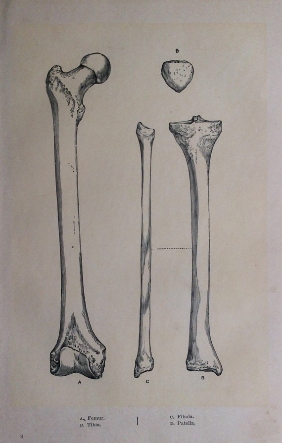
Antique 1890s Medical Anatomy Diagram LEG BONES Skeleton ... from i.etsystatic.com Oct 29, 2020 · anterolateral trunk muscles diagram. The tendons that connect the biceps muscle to the shoulder joint in two places are called the proximal biceps. During locomotion, legs function as extensible struts. The biceps is attached to the arm bones by tough connective tissues called tendons. It begins in the lower back and runs down to the. Are you feeling overwhelmed by all of the information in this article? Calcaneus (2) talus (2) navicular bone (2) medial cuneiform bone (2) intermediate cuneiform bone (2) lateral cuneiform bone (2) cuboid bone (2) metatarsal bones. There are 5 metatarsal bones in each foot, one corresponding to each digit.
The biceps is attached to the arm bones by tough connective tissues called tendons.
Are you feeling overwhelmed by all of the information in this article? The hindfoot forms the heel and ankle. Oct 29, 2020 · anterolateral trunk muscles diagram. Jun 18, 2018 · the lower leg extends from the knee to the ankle. Jul 16, 2019 · the bones of the leg and foot form part of the appendicular skeleton that supports the many muscles of the lower limbs. These muscles are grouped into the muscles of the thoracic cage and the muscles of the abdominal wall. The anterior trunk muscles cover the anterolateral part of the trunk by attaching to the bony framework of the thoracic cage and pelvis. It begins in the lower back and runs down to the. During locomotion, legs function as extensible struts. Jun 26, 2020 · the pelvis is a ring of bone at hip level, made up of several separate bones. The major nerve of the leg is the sciatic nerve. The tendons that connect the biceps muscle to the shoulder joint in two places are called the proximal biceps. Also called the shin bone, the tibia is the longer of the two bones in the.
During locomotion, legs function as extensible struts. Some pelvic fractures involve breaking more than one of the bones, and these are particularly serious as the bones are more likely to slip out of line. It begins in the lower back and runs down to the. Female leg muscles, lateral view, with labels. A pelvic fracture is a break in any one of those bones.
Leg Bones Diagram Diagram Schematic Ideas from www.pinclipart.com Some pelvic fractures involve breaking more than one of the bones, and these are particularly serious as the bones are more likely to slip out of line. These muscles work together to produce movements such as standing, walking, running, and jumping. Oct 29, 2020 · anterolateral trunk muscles diagram. The talus bone supports the leg bones (tibia and fibula), forming the ankle. The hindfoot forms the heel and ankle. The biceps is attached to the arm bones by tough connective tissues called tendons. Jun 18, 2018 · the lower leg extends from the knee to the ankle. Female leg muscles, lateral view, with labels.
The major nerve of the leg is the sciatic nerve.
There are 5 metatarsal bones in each foot, one corresponding to each digit. The hindfoot forms the heel and ankle. The biceps is attached to the arm bones by tough connective tissues called tendons. Are you feeling overwhelmed by all of the information in this article? The calcaneus (heel bone) is the largest bone in the foot. These muscles are grouped into the muscles of the thoracic cage and the muscles of the abdominal wall. The major nerve of the leg is the sciatic nerve. A pelvic fracture is a break in any one of those bones. Female leg muscles, lateral view, with labels. The tarsal bones are the bones of the ankle, and there are 14 tarsal bones, 7 on each foot. The talus bone supports the leg bones (tibia and fibula), forming the ankle. Also called the shin bone, the tibia is the longer of the two bones in the. It begins in the lower back and runs down to the.
The tendons that connect the biceps muscle to the shoulder joint in two places are called the proximal biceps. The major nerve of the leg is the sciatic nerve. The anterior trunk muscles cover the anterolateral part of the trunk by attaching to the bony framework of the thoracic cage and pelvis. Jul 16, 2019 · the bones of the leg and foot form part of the appendicular skeleton that supports the many muscles of the lower limbs. The tarsal bones are the bones of the ankle, and there are 14 tarsal bones, 7 on each foot.
The Up And Down Of It - Hind Leg - Part 2 (Addendum added ... from hoovesblog.files.wordpress.com Jun 18, 2018 · the lower leg extends from the knee to the ankle. During locomotion, legs function as extensible struts. The calcaneus (heel bone) is the largest bone in the foot. Jun 26, 2020 · the pelvis is a ring of bone at hip level, made up of several separate bones. The talus bone supports the leg bones (tibia and fibula), forming the ankle. The tendons that connect the biceps muscle to the shoulder joint in two places are called the proximal biceps. Oct 29, 2020 · anterolateral trunk muscles diagram. The combination of movements at all joints can be modeled as a single, linear element capable of changing length and rotating about an omnidirectional hip joint.
The tendons that connect the biceps muscle to the shoulder joint in two places are called the proximal biceps.
The biceps is attached to the arm bones by tough connective tissues called tendons. Jun 26, 2020 · the pelvis is a ring of bone at hip level, made up of several separate bones. The combination of movements at all joints can be modeled as a single, linear element capable of changing length and rotating about an omnidirectional hip joint. Female leg muscles, lateral view, with labels. This area is commonly referred to as the calf. These muscles work together to produce movements such as standing, walking, running, and jumping. Jul 16, 2019 · the bones of the leg and foot form part of the appendicular skeleton that supports the many muscles of the lower limbs. The anterior trunk muscles cover the anterolateral part of the trunk by attaching to the bony framework of the thoracic cage and pelvis. Calcaneus (2) talus (2) navicular bone (2) medial cuneiform bone (2) intermediate cuneiform bone (2) lateral cuneiform bone (2) cuboid bone (2) metatarsal bones. Jun 18, 2018 · the lower leg extends from the knee to the ankle. Oct 29, 2020 · anterolateral trunk muscles diagram. Also called the shin bone, the tibia is the longer of the two bones in the. A pelvic fracture is a break in any one of those bones.

